
Keyhole joint surgery: what the camera can and cannot do
A camera the width of a pencil goes into the joint through a cut a few millimetres long, and the joint is filled with fluid so the surgeon can see. It changed what is possible inside a knee or a shoulder — and the small scars are the least interesting thing about it.
What it actually is
Arthroscopy (arthroscopy) means looking inside a joint with a camera. The word is literally that: arthro, joint, and scopy, to look.
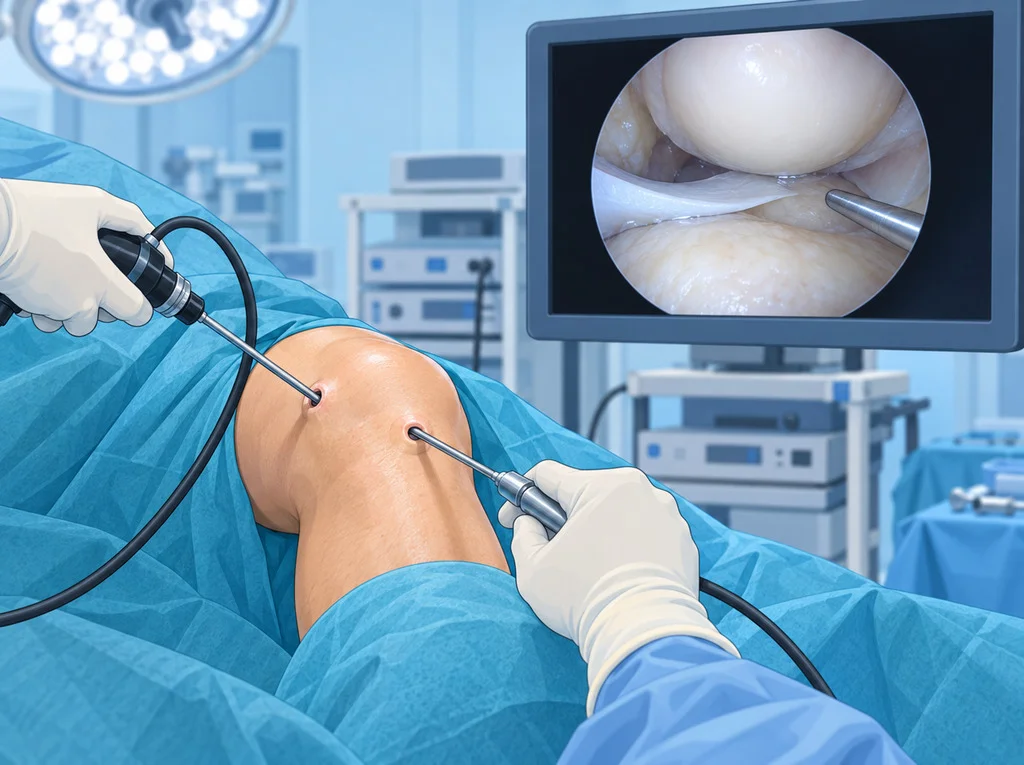
A rod about the width of a pencil, carrying a lens and a light, goes in through a cut a few millimetres long. What it sees appears, magnified, on a screen. A second small cut lets the surgeon pass instruments in alongside it, and a third is added when one is needed.
Before this existed, seeing inside a knee meant opening it. That difference is the point of the whole technique — not the cosmetics of the scar, but that a surgeon can now look at, and work on, the inside of a joint without taking it apart to get there.
Why the joint is filled with fluid
This is the part nobody expects, and it explains a lot about the operation.
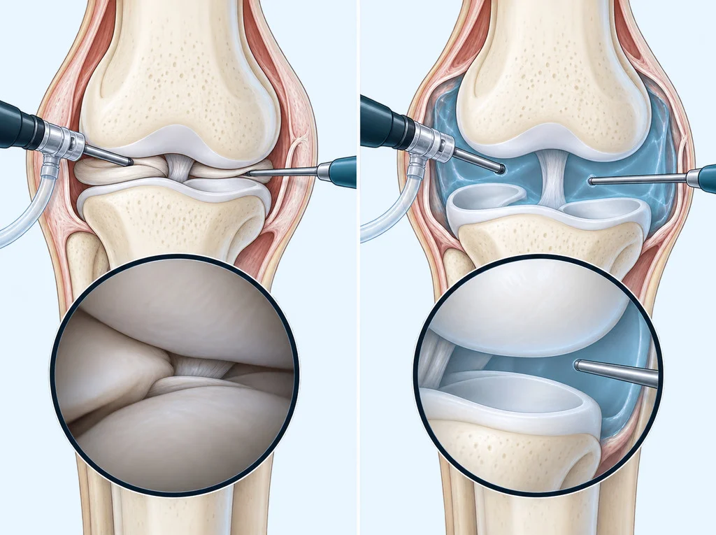
A joint is not a room with air in it. The surfaces sit against each other, with only a film of fluid between. Put a camera into that and you see nothing at all — the lens is pressed against tissue.
So sterile fluid is run into the joint throughout the operation. It pushes the space open, keeps the view clear by washing blood away, and lets instruments move. It is also why:
- the joint feels tight and swollen afterwards. Some of that fluid stays behind for a day or two;
- a dressing can leak in the first hours, which is expected rather than alarming;
- the operation has a time pressure that an open operation does not — fluid under pressure gradually seeps into the tissue around the joint.
What it is good at
The camera is a way in. What matters is what is done once you are in there.
- Looking. Sometimes the honest answer is that the scan, the examination and the story do not agree, and a direct look settles it. This is much rarer than it used to be, because MRI got good.
- Removing what should not be there. A torn flap catching in the joint, a loose fragment, inflamed lining.
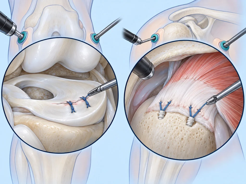
- Repairing torn tissue. Stitching a torn meniscus back down, re-attaching a torn rotator cuff tendon to bone.
- Rebuilding. A new cruciate ligament can be placed through the same small openings, using tunnels drilled in the bone.
- Making room. Trimming bone that is catching against tendon or against another bone.
What it cannot do
This section matters more than the one above it, because the picture most people have of keyhole surgery is of a general-purpose repair.
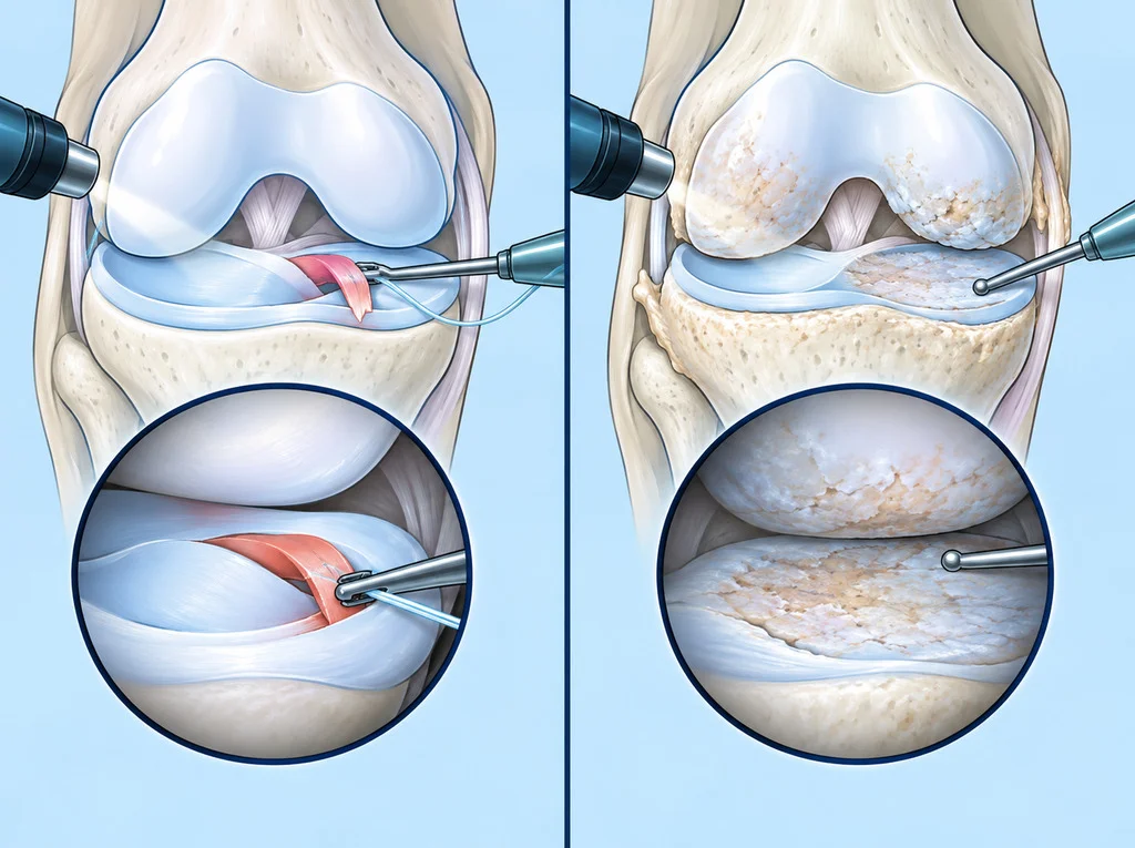
It cannot grow back a worn joint surface. Once articular cartilage has thinned, no instrument restores it. The camera can smooth a flap that is catching; it cannot resurface the joint.
That is not a small caveat. An international guideline panel reviewing arthroscopy for degenerative knee disease — wear, with or without a meniscal tear found on a scan — recommended against it for almost all such patients, because the benefit does not last and the alternative is not nothing. Systematic reviews comparing a proper exercise programme with arthroscopic surgery for degenerative meniscal tears have repeatedly found similar results for pain and function.
This is also the general pattern across musculoskeletal care: reviews of high-quality guidelines consistently put exercise, education and load management first, and reserve surgery for the cases where it is genuinely the thing that helps.
Small cuts do not mean a small operation
If you take one thing from this page, take this.
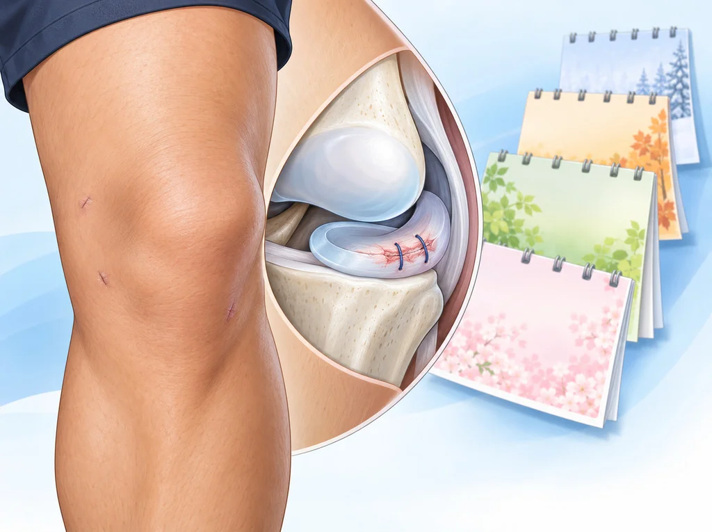
The scars from an arthroscopy are a few millimetres each and are healed in a couple of weeks. What was done inside is on a completely different timetable.
- Removing a loose fragment: the joint may settle within weeks.
- Repairing a meniscus: the stitched tissue needs months, and there are usually limits on bending and weight-bearing while it heals.
- Reconstructing a ligament, or repairing a tendon to bone: the new tissue has to grow into the bone. That is a matter of months, with a staged rehabilitation programme, and rushing it is how repairs fail.
A skin wound that looks finished is not evidence that the inside is. This is why the rehabilitation plan, not the appearance of the leg, is what tells you where you are — and why “keyhole” and “quick” are not synonyms.
What the day is like
The details vary by hospital, joint and operation, and the plan you are given is the one that applies to you. In general terms:
- It is usually planned as day surgery. In a registry analysis of almost 100,000 day-case knee, hip and shoulder arthroscopies, about 2.6% were admitted overnight rather than going home.
- Anaesthetic may be general, spinal, a regional block, or a combination. The anaesthetist will go through it with you.
- You will wake with a bandaged joint, and often a swollen one.
- Someone should take you home, and you should not drive that day.
- Pain relief, a plan for moving, and the date physiotherapy starts should all be given to you before you leave.
Whether to have the operation at all — how to weigh it, what to ask, what happens if you wait — is a separate question, and the treatment decisions page is about exactly that.
When to see a doctor
Every operation carries risk, including infection, a clot in the leg or lung, stiffness, and nerve or vessel injury. These are uncommon after arthroscopy, but they are not zero, and the person taking your consent should go through the ones that apply to your operation specifically.
Questions to ask your doctor
- What exactly do you expect to do inside the joint, and what would change that during the operation?
- What is this operation expected to improve — the pain, the locking, the stability, or something else?
- What happens if I wait, or do a rehabilitation programme first?
- How long until I can walk normally, drive, work and go back to sport?
- What are the risks of this particular operation for someone like me?
Frequently asked questions
Small cuts must mean an easy recovery — is that right?
No, and it is the most common misunderstanding about this operation. The skin heals in a couple of weeks whatever was done underneath. How long the recovery takes depends on what was repaired inside and how long that tissue needs — a repaired ligament or tendon is on its own timetable and does not care how small the scars are.
Can the camera fix worn cartilage?
It can see it, and it can tidy loose flaps that are catching. It cannot grow a worn joint surface back. Guidelines on arthroscopy for a degenerative knee are clear that it does not give a lasting benefit for pain from wear itself, and exercise and weight management are the treatments with the evidence behind them.
Is it done under general anaesthetic?
That depends on the joint, the operation and you — general, spinal and regional blocks are all used, often in combination. The anaesthetist discusses it with you beforehand, and it is a reasonable thing to ask about early if it worries you.
Will I stay in hospital?
Most joint arthroscopy is planned as day surgery, and most people do go home the same day. In one registry analysis of nearly 100,000 day-case knee, hip and shoulder arthroscopies, about 2.6% were admitted. Your own plan depends on the operation, the anaesthetic and your other health.
References
- Siemieniuk RAC et al. Arthroscopic surgery for degenerative knee arthritis and meniscal tears: a clinical practice guideline. BMJ. 2017
- Giuffrida A et al. Conservative vs. surgical approach for degenerative meniscal injuries: a systematic review of clinical evidence. Eur Rev Med Pharmacol Sci. 2020
- Gabriel RA et al. The association of body mass index with same-day hospital admission, postoperative complications, and 30-day readmission following day-case eligible joint arthroscopy: A national registry analysis. J Clin Anesth. 2019
- Lin I et al. What does best practice care for musculoskeletal pain look like? Eleven consistent recommendations from high-quality clinical practice guidelines: systematic review. Br J Sports Med. 2019
Related conditions


Written by Assoc. Prof. Sorawut Thamyongkit, M.D.