
What a knee MRI shows
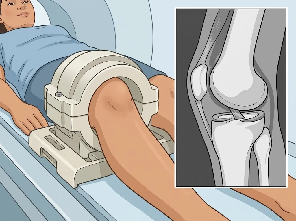
A knee MRI shows the menisci, the cruciate ligaments, cartilage and swelling in bone. What it finds has to be read against your symptoms, because knees that never hurt show abnormalities almost every time.
What is being looked at
In a knee MRI the radiologist works through the main structures:
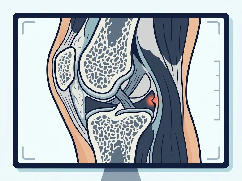
- The menisci — the two C-shaped pads of cartilage that spread load. Is there a tear, what kind, and where?
- The anterior and posterior cruciate ligaments — is each one continuous?
- The collateral ligaments on the inner and outer sides
- Joint cartilage — thinned, fissured, or lost
- Bone marrow oedema, which usually means force or abnormal loading at that spot
- Fluid in the joint and in the surrounding bursae
- The patellar and quadriceps tendons
What to understand before reading the result
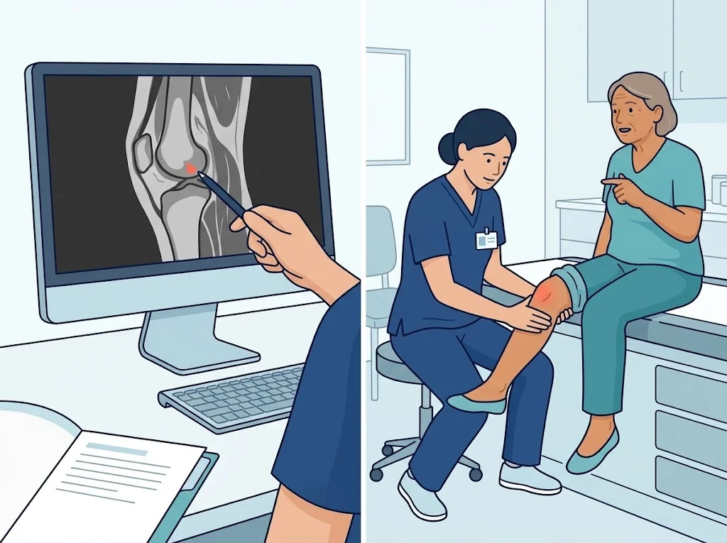
This is why a doctor will ask about your symptoms in detail and examine the knee by hand before deciding whether what is on the scan is relevant. The question being answered for you is not “what is on the scan” but “what on the scan explains your symptoms”.
Words in a knee MRI report
| In the report | In plain language |
|---|---|
| meniscal tear | a tear in a meniscus; there are several patterns — horizontal, radial, complex |
| degenerative tear | a tear from wear over time rather than a single injury |
| ACL / PCL intact | the cruciate ligament is continuous |
| chondral thinning | the joint cartilage is thinner than expected |
| bone marrow oedema | fluid within the bone, usually from impact or abnormal loading |
| joint effusion | more fluid in the joint than normal |
| Baker cyst | a fluid-filled swelling behind the knee, often alongside a problem inside the joint |
So is surgery needed?
That depends on much more than the pictures. A systematic review comparing arthroscopic surgery with non-surgical management in degenerative meniscal tears found that, in people without locking or catching, a proper course of physiotherapy gave results for pain and function comparable to surgery, and recommended starting with non-surgical treatment.
Situations where surgery tends to be considered sooner include a knee locked so that it will not straighten or bend, a tear at the root of the meniscus, or an ACL injury in someone who needs to return to pivoting sport. These are decisions to make with your surgeon.
Questions to ask your doctor
- On my scan, which findings are likely to be causing symptoms, and which are common for my age?
- If I choose not to have surgery, what is the plan, and by when should I see a change?
- What symptoms mean I should come back before my next appointment?
- What are the advantages and disadvantages of surgery in my case?
- What activities should I avoid for now?
Frequently asked questions
The report says a meniscal tear. Do I need surgery?
Not necessarily. Degenerative tears are common in older knees with no symptoms, and a systematic review found that for degenerative tears without locking, physiotherapy gave results comparable to arthroscopic surgery. The decision depends on the type of tear, your age and your symptoms.
Should every painful knee be scanned?
No. Many knee problems are diagnosed from the history and examination. MRI is usually ordered when a ligament or meniscal injury is suspected, when symptoms are not settling as expected, or when surgery is being planned.
Can an MRI grade my osteoarthritis?
MRI shows cartilage in more detail than an X-ray, but in practice osteoarthritis is staged using X-rays alongside symptoms. Your doctor picks the test that answers the question being asked.
References
Related conditions




Written by Assoc. Prof. Sorawut Thamyongkit, M.D.