
Musculoskeletal ultrasound
Ultrasound shows tendons and the soft tissue around a joint in minutes, can watch them while you move, and uses no radiation. It also depends heavily on the operator, and cannot see through bone.
How it works
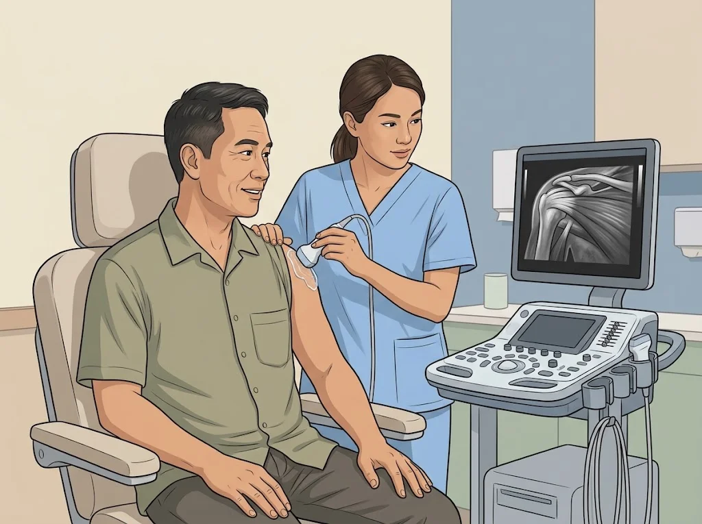
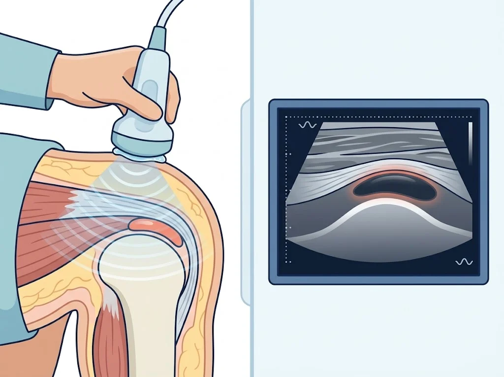
The probe sends high-frequency sound into the tissue and listens for the echo. Tissues of different density reflect differently, and the machine builds a live image — moving, not a still.
The gel exists to push air out from between the probe and your skin, because air blocks almost all of the sound.
What it is good at
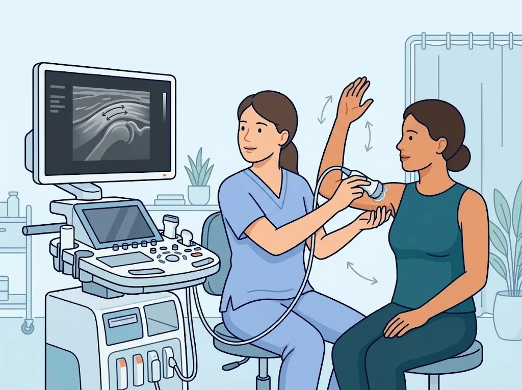
Watching movement. This is the clearest advantage. The doctor can ask you to lift your arm and watch whether a tendon glides normally through its space, or ask you to bend a finger and see exactly where the tendon catches. MRI cannot do this — it is a still image in one position.
Speed, and no radiation. A scan takes minutes, can be repeated, and both sides can easily be compared in one sitting.
Guiding a needle. When fluid is being drawn off or an injection placed, ultrasound lets the doctor see the needle tip as it goes.
Accuracy for some questions. A meta-analysis of ultrasound for rotator cuff tears found a pooled how well a test picks up people who really do have the condition; higher means fewer missed of about 95% and a pooled how well a test correctly clears people who do not have the condition; higher means fewer false alarms of about 72% against arthroscopy or MRI. Sensitivity that high makes it useful for ruling a tear out. In emergencies, another meta-analysis found bedside ultrasound highly accurate for diagnosing a dislocated shoulder.
A test with high sensitivity but lower specificity misses few real cases but may report an abnormality in someone who does not have one.
Its limits
- It cannot see through bone. Anything deep inside a joint, or shadowed by bone, is not visible.
- It depends on the operator. Two examiners can report differently. This is the nature of the test, not a failing of either.
- It does not show swelling within bone, which MRI does.
- Deep structures — a hip in someone with more soft tissue, for instance — can be hard to see clearly.
Where it is used often
- Rotator cuff tears and tendon inflammation
- The Achilles tendon and the plantar fascia
- Trigger finger, watching the tendon as the finger bends and straightens
- Bursae around joints, and fluid within a joint
- Guiding injections and aspirations
Questions to ask your doctor
- What question will this scan answer about my symptoms?
- Is the result enough to plan treatment, or will something else be needed?
- If the ultrasound does not match my symptoms, what happens next?
- Will this need to be repeated later?
Frequently asked questions
Can ultrasound replace MRI?
For some questions, yes — it is very sensitive for rotator cuff tears, for example. For questions that need to see deep inside a joint or within bone, MRI is still needed. Your doctor chooses based on what needs to be known.
Why did two hospitals report different things?
Ultrasound depends on the operator more than most tests — the angle of the probe, your position, and experience all matter. Different reports can both be honest work.
Does it hurt, and how do I prepare?
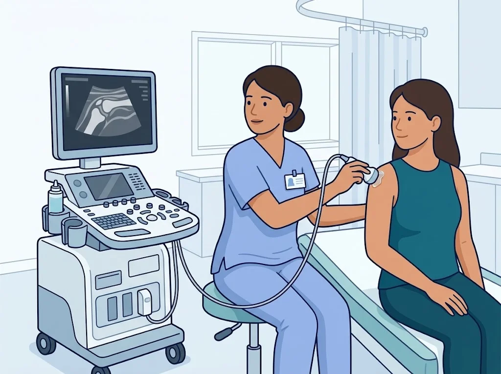
It does not hurt. Gel is applied and a probe is moved over the skin, sometimes pressing to see a structure better. No fasting is needed. Wear clothes that make the area easy to reach.
References
Related conditions






Written by Assoc. Prof. Sorawut Thamyongkit, M.D.