
What an MRI scan is
MRI shows ligaments, cartilage and soft tissue in far more detail than an X-ray. That same detail means it finds things that are not causing your symptoms. This page covers both sides.
How it works
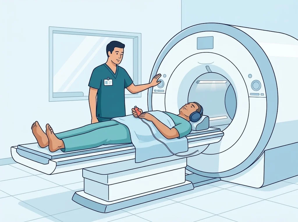
MRI stands for magnetic resonance imaging. The machine creates a very strong magnetic field and sends radio waves through it. Water molecules in different tissues respond differently, and a computer assembles the returning signals into cross-sectional images, slice by slice.
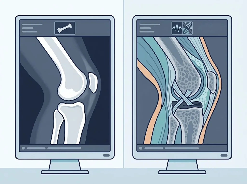
Because soft tissues differ in water content, MRI can tell ligament from muscle from meniscus from cartilage — which an X-ray cannot do.
What it shows that an X-ray does not
- Ligaments such as the ACL, and tendons such as the rotator cuff
- The menisci of the knee, and the discs of the spine
- Joint cartilage directly
- fluid within the bone under a joint surface, often after injury or unusual loading, which is sometimes an important clue
- Inflammation, masses and infection at a stage where an X-ray is still normal
The most important limitation — it finds too much
This is worth knowing before you go.
That is why systematic reviews of high-quality guidelines recommend using imaging selectively — when the result will change a decision, rather than for reassurance.
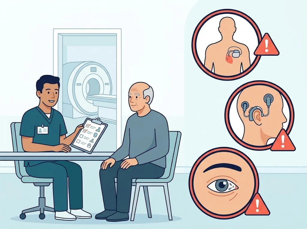
What to declare before you go in
Also mention a joint replacement, plates or screws, large or very recent tattoos, medication patches, or pregnancy. Most modern metal implants are safe in an MRI scanner, but that is a judgement for the staff to make, not something to guess at.
What the scan is like
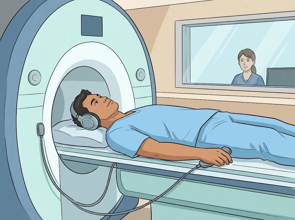
You lie on a table that slides into the tube, with the body part being scanned at the centre of the magnet. The machine makes loud rhythmic knocking sounds. You will be given ear protection and a call button.
The most important thing is to stay still. Even small movements blur the images enough to make them unreadable, and the sequence has to be repeated.
If small spaces frighten you, say so when the scan is booked rather than on the day. There is a range of things that help, from a careful explanation of what will happen through to medication if your doctor thinks it appropriate.
Questions to ask your doctor
- Why this scan now, and how will the result change the plan?
- Is what was found the cause of my symptoms, or something common for my age?
- If I do not have the scan now, what are the alternatives?
- Do I need contrast, and why?
- I have metal or a device implanted — what do I need to do?
Frequently asked questions
Does an MRI hurt?
No, but you have to lie still inside a narrow tube for 20 to 60 minutes, and the machine is very loud. You will be given earplugs or headphones. If small spaces frighten you, say so in advance so it can be planned for.
Is there radiation?
No. MRI uses a magnetic field and radio waves, not X-rays. Its safety questions are about metal and implanted devices, not about dose.
The report shows a tear. Do I need an operation?
Not necessarily. Many of the findings on a scan are also present in people with no symptoms at all. Treatment decisions come from your symptoms, the examination, and how you respond to non-surgical care together — not from the images alone.
Why do some people need an injection during the scan?
Sometimes a contrast agent is used to make inflammation, a lump, or the inside of a joint stand out. Your doctor will explain why and will ask about allergies and kidney function first.
References
- Horga LM et al. Prevalence of abnormal findings in 230 knees of asymptomatic adults using 3.0 T MRI. Skeletal Radiol. 2020
- Lin I et al. What does best practice care for musculoskeletal pain look like? Eleven consistent recommendations from high-quality clinical practice guidelines: systematic review. Br J Sports Med. 2019
Related conditions






Written by Assoc. Prof. Sorawut Thamyongkit, M.D.