
X-rays of bones and joints
An X-ray shows bone clearly and soft tissue barely at all. This page explains what it can answer, what it cannot, and why a film that looks bad does not mean your symptoms will be bad too.
What an X-ray is
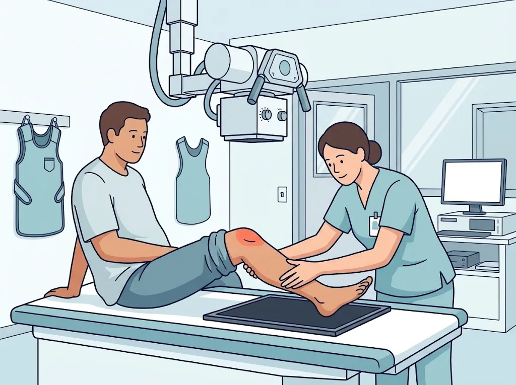
An X-ray passes a small amount of radiation through the body onto a detector. Different tissues let it through to different degrees. Bone, which is full of calcium, blocks most of it and appears white; muscle and fat let more through and come out grey to black.
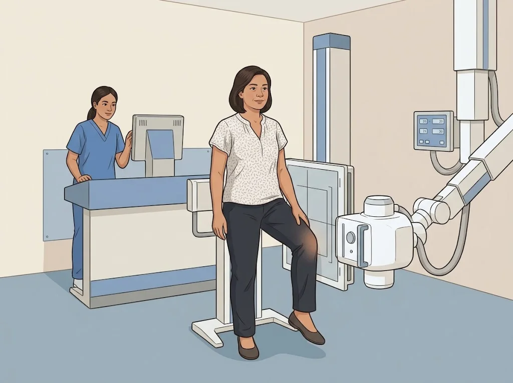
The picture takes seconds, does not hurt, and the whole visit is usually a few minutes. You will be asked to hold a position and stay still, and more than one view is often taken — a three-dimensional structure squashed into a single flat image can hide things.
What an X-ray can answer
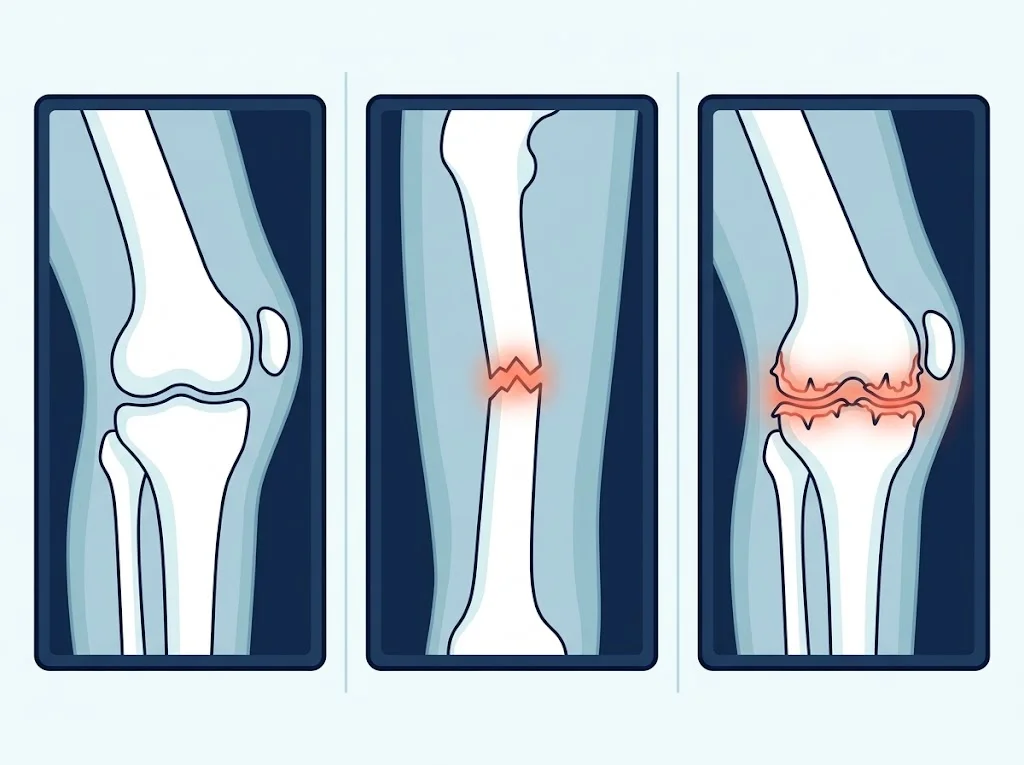
- Fractures, especially where the bone has moved out of place
- Dislocations — whether the bone is still in its socket
- A narrowed joint space, which is indirect evidence that the the smooth surface covering the ends of bones inside a joint, letting it glide and absorb shock has thinned
- Bone spurs at the joint margins, seen in osteoarthritis
- Alignment — bow legs, or a curved spine
- Lumps or abnormal areas within bone that need further study
What an X-ray cannot show
This is where most confusion starts. An X-ray does not clearly show:
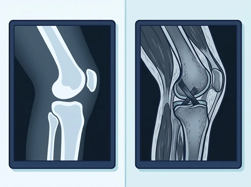
- ligaments and tendons, such as the ACL or the rotator cuff
- the menisci of the knee
- joint cartilage directly (only indirectly, through the joint space)
- nerves and spinal discs
- early inflammation or fluid in a joint
So a “normal” X-ray does not mean nothing is wrong. It means nothing was found among the things an X-ray can see.
Why your doctor may not order one
A systematic review of high-quality clinical practice guidelines for musculoskeletal pain distilled eleven recommendations they all agree on. One of them is to use imaging selectively, rather than scanning everyone.
The reason is not only cost. Finding a small abnormality that is not causing your symptoms can lead to more tests, more worry, and treatment nobody needed. For many conditions the history and examination tell a doctor more than people expect.
Imaging is usually ordered when a fracture or dislocation is suspected, after significant trauma, when symptoms are not improving as they should, or when a treatment is being planned that depends on the state of the bone.
Getting ready
- Wear something comfortable, without metal buttons, zips or hooks over the area
- Take off jewellery near the area
- Tell the radiographer if you are, or might be, pregnant
- Bring any previous films or discs. Comparing with an older image is worth a great deal
Reading the report
The report is written by a radiologist for the doctor who requested it — not for you. Common phrases:
| In the report | In plain language |
|---|---|
| joint space narrowing | the gap between two bones is reduced, usually meaning thinner cartilage |
| osteophyte / spur | a bony outgrowth at the joint edge, seen in osteoarthritis |
| subchondral sclerosis | denser bone under the joint surface, an adaptation to load |
| no acute fracture | no new break was seen |
| degenerative change | changes that come with use and age |
| soft tissue swelling | the tissues around the joint are swollen |
These describe what is visible. They do not say how much you hurt or what should be done. Treat the report as one piece of information and ask your doctor what it means in your case.
Questions to ask your doctor
- Will this X-ray change my treatment, and how?
- Does what you see on the film explain all of my symptoms?
- Do I need any other test, and why?
- Should this be repeated, and if so when?
- Can I have a copy of the images?
Frequently asked questions
Is the radiation dose safe?
The dose for a limb or a joint is very low. Your doctor orders one when the result is likely to change what happens next. If you are worried, or if you are or might be pregnant, tell the radiographer before you go in — always.
My knee hurts a lot but my doctor did not order an X-ray. Is that wrong?
Not necessarily. High-quality guidelines agree that imaging should be used selectively. For many problems the history and examination give more information than a film, and starting treatment works just as well. If you want to understand the reasoning, ask directly.
The report says osteoarthritis. Does that mean surgery?
No. How worn a joint looks on a film and how much it hurts do not track each other closely. Treatment decisions rest on your symptoms, how you live, and how you respond to non-surgical care — not on the film alone.
References
- Lin I et al. What does best practice care for musculoskeletal pain look like? Eleven consistent recommendations from high-quality clinical practice guidelines: systematic review. Br J Sports Med. 2019
- French HP et al. Prevalence of neuropathic pain in knee or hip osteoarthritis: A systematic review and meta-analysis. Semin Arthritis Rheum. 2017
Related conditions




Written by Assoc. Prof. Sorawut Thamyongkit, M.D.